Fetal Echocardiography / Your Unborn Baby's Heart
Overview of congenital heart disease
Congenital heart disease is a problem that occurs with the baby's heart while the baby is still developing. It's seen in approximately 1% of babies born in the United States and is the most common form of birth defect.
The baby's heart begins to form immediately after conception and is complete by eight week's gestation. The heart begins as a tube shaped structure that twists and divides to form the heart and heart valves. A congenital heart defect usually occurs because the heart does not twist or divide normally. Some mothers wonder if drugs, alcohol or medications contributed to their child's heart defect. In most cases we don't know why these defects occur. Although, some heart defects can run in families or be related to a disease the mother has, diabetes mellitus, for example.
Congenital heart defects range from mild to very severe. Some require surgical repair in the newborn period and some may resolve on their own with time. Your pediatric cardiologist will be able to counsel you as to the severity of your baby's heart defect.
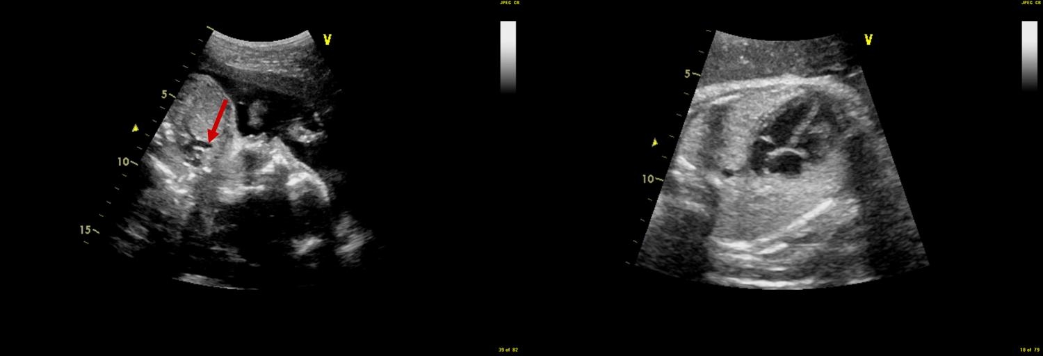
What is fetal echocardiogram?
A fetal echocardiogram is a detailed ultrasound performed of the baby's heart before the baby is born. A small camera called a transducer is placed on the pregnant mother's abdomen and sends out ultrasonic sound waves. The ultrasound waves bounce off the baby's organs, including the heart and are sent back to the camera which then creates a moving picture of the different parts of the heart for the doctor to evaluate. The sound waves can also detect blood flow throughout the baby's heart. This enables the doctor to evaluate the structure and function of the fetal heart.
Who needs one?
Fetal echocardiograms are recommended for the following women:
- If a first degree relative has been diagnosed with a congenital heart defect. First degree relative includes the mother or father of the baby as well as any siblings of the baby
- If there is a known family history of disorders that are passed along from generation to generation such as Marfan's syndrome or tuberous sclerosis
- If the unborn baby has been diagnosed with a genetic abnormality including disorders with an abnormal number of chromosomes; Down syndrome, for example
- Abnormal amniocentesis
- If the mother has taken medications that are known to cause congenital heart defects, Accutane, for example.
- If the mother has specific health problems such as diabetes (the type that the mother had prior to pregnancy), phenylketonuria or autoimmune diseases such as systemic lupus erythematosis
- If the mother had specific infections during pregnancy such as rubella or CMV
- If a heart abnormality is suspected on routine ultrasound
- If there are abnormalities outside of the heart of the fetus noted on routine prenatal ultrasound; examples include extra fluid around the lungs or the heart or an abnormality of another organ such as the kidneys or brain.
- Abnormal fetal heart rate or rhythm. This can be an irregular heart beat or heart rate that is too fast or too slow.


